Dental implants have a strong track record, but failures still happen and can lead to costly repairs and patient disappointment.
Advanced imaging technologies and digital planning tools significantly reduce the risk of dental implant failure by allowing dentists to identify potential problems before surgery and place implants with greater precision.
When you combine detailed 3D scans with computer-guided planning, your dentist can avoid critical structures, ensure proper implant positioning, and select the right implant size for your specific anatomy.
Digital planning minimizes the chance of injury or improper fit, and surgical guides produced from these digital models further enhance precision during your actual procedure. You benefit from this heightened accuracy with fewer surgical surprises, less discomfort, and lower risk of complications.
The difference between traditional methods and modern imaging approaches can mean the difference between an implant that lasts decades and one that fails within months.
Understanding how these technologies work together to protect your investment in dental implants helps you make informed decisions about your treatment. From initial scans to final placement, each step in the modern implant process is designed to give you the best possible outcome.
Key Takeaways
- Advanced 3D imaging and digital planning allow dentists to identify risks and place implants more accurately before surgery begins
- Computer-guided surgical techniques reduce complications and improve implant success rates compared to traditional methods
- Better planning leads to fewer surprises during surgery, faster healing times, and implants that last longer
Get advanced CBCT guided implant planning in Munster and Schererville, IN. Schedule your consultation today.
Understanding Implant Failure Risks
Dental implants can fail for reasons ranging from infections and poor bone integration to patient health conditions and surgical planning mistakes. Lack of osseointegration accounts for 36.4% of failures, while absence of primary stability causes 22.4% of cases.
Common Causes of Dental Implant Failure
Dental implant failure happens when your implant doesn’t integrate properly with your jawbone or develops problems after placement. The most common issue is poor osseointegration, which means the bone doesn’t fuse correctly with the implant surface.
Peri-implantitis represents 14% of implant failures and occurs when infection develops around the implant. This condition causes inflammation and peri-implant bone loss that weakens the implant’s foundation.
Infections account for 7.5% of failures and can happen early or late in the healing process. Early failures typically occur before your implant heals completely. Late failures happen after your implant has already integrated but develops peri-implant disease over time.
Other causes include:
- Excessive force on the implant from grinding or clenching
- Improper implant positioning that makes restoration difficult
- Trauma to the implant site
- Inadequate primary stability during placement
The Impact of Poor Planning on Implant Outcomes
Poor implant placement and inadequate bone quality significantly increase your risk of complications. When your dentist doesn’t properly assess bone density or volume before surgery, the implant may not have enough support to succeed.
Iatrogenic issues related to incorrect location or position cause 14% of failures. These problems happen when implants are placed at wrong angles or depths. Without proper planning, your implant might end up too close to nerves, sinuses, or adjacent teeth.
Bone grafting requirements often go unnoticed without thorough evaluation. About 30.2% of failed implant cases involved previous bone grafting procedures. When your dentist skips detailed imaging, they might miss areas where bone augmentation is necessary.
Implant length and width selection matters too. Most failures occur with 10mm implants, which represent 63.4% of cases. Your specific anatomy requires precise measurements to choose the right implant dimensions.
Patient-Related Factors Affecting Success
Your overall health plays a major role in implant success. Smokers, people with uncontrolled diabetes, and those with osteoporosis face higher failure risks.
Medical conditions affect 34.1% of implant failure cases. The most common health issues include:
| Condition | Percentage |
| Hypertension | 15.2% |
| Controlled diabetes | 9.8% |
| Hypothyroidism | 4.5% |
| Osteoporosis | 3.0% |
Your bone quality directly impacts osseointegration. Osteoporosis weakens your jawbone and makes it harder for implants to integrate properly. Bisphosphonate medications for bone conditions appear in 2.3% of failure cases.
Periodontal disease affects nearly half of all patients who experience failures. If you have active gum disease, bacteria can spread to your implant site and cause peri-implantitis. Poor oral hygiene increases your risk of developing peri-implant disease.
Smoking reduces blood flow to your gums and slows healing. Gender also influences outcomes, with significant differences in failure mechanisms between males and females.
Want lower risk and better precision? Book a Munster or Schererville, IN implant planning visit with a surgical team.
The Critical Role of Imaging in Implant Success
Proper imaging allows your dental team to see bone structure, nerve locations, and potential complications before surgery begins. Different imaging methods offer varying levels of detail, with newer technologies providing three-dimensional views that transform how dentists plan your implant placement.
Advancements in Digital Imaging Technologies
Digital imaging has changed how dentists plan dental implants. Traditional two-dimensional x-rays only show flat images of your jaw, which can miss important details about bone depth and nerve positions.
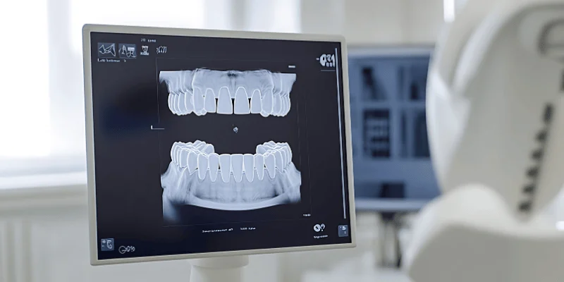
3D imaging technology has revolutionized dental implant surgery by giving dentists a complete view of your mouth and jaw structure. Cone beam computed tomography (CBCT) creates detailed three-dimensional scans that show your bone from every angle.
This helps your dentist see exactly where nerves run through your jaw and measure bone thickness with precision.
Computer-aided design and manufacturing (CAD/CAM) systems work with these scans to create surgical guides. These guides help your dentist place implants in the exact position planned on the computer. Digital planning minimizes the chance of injury or improper fit, which means fewer problems during and after your surgery.
Comparing CBCT, Panoramic, and Periapical Imaging
Different imaging methods give dentists different types of information about your mouth.
Panoramic radiographs capture your entire mouth in one flat image. These panoramic x-rays show all your teeth, both jaws, and surrounding structures. They’re useful for getting a general overview but don’t provide detailed measurements of bone thickness.
Periapical radiographs focus on specific teeth and the bone around them. Your dentist uses these to check individual tooth roots and nearby bone. However, periapical radiographs only show two dimensions.
Cone beam computed tomography provides the most detailed view. CBCT scans create hundreds of images that combine into a 3D model of your jaw. You can measure bone height, width, and density accurately with computed tomography. CBCT imaging helps avoid nerve and sinus damage, reducing complications during your surgery.
| Imaging Type | Dimensions | Best For | Limitations |
| Panoramic | 2D | Overall view | No depth measurement |
| Periapical | 2D | Individual teeth | Limited area coverage |
| CBCT | 3D | Implant planning | Higher cost |
Imaging for Bone Assessment and Nerve Mapping
Your dentist needs to know if you have enough bone to support an implant. Imaging shows both bone quality and bone quantity in your jaw.
CBCT scans measure your alveolar bone in three dimensions. This tells your dentist if the bone is thick enough and tall enough for an implant. Areas with thin bone might need grafting before implant placement.
Bone quality matters as much as bone thickness. Dense bone holds implants better than soft bone. Your dentist can assess bone density through imaging to predict how well your implant will integrate.
Diagnostic imaging helps identify potential issues before surgery. Nerve mapping shows exactly where the inferior alveolar nerve runs through your lower jaw. Damaging this nerve during surgery can cause numbness in your lip and chin. Upper jaw scans reveal sinus locations to prevent penetration during implant placement.
Reducing Radiation Risks with Modern Techniques
Modern imaging equipment uses less radiation than older systems. CBCT machines have improved to deliver detailed scans with lower radiation doses.
Your dentist only orders imaging when necessary based on your examination. Not every patient needs the same imaging. Some cases require only panoramic radiography, while complex cases benefit from cone beam computed tomography.
Newer CBCT units have adjustable settings that limit the scan area to just what’s needed. Scanning only the implant site instead of your entire jaw reduces your radiation exposure. The machines also use faster sensors that capture images quickly, shortening scan times.
Protective equipment like lead aprons shield the rest of your body during imaging. Digital sensors are more sensitive than old film, requiring less radiation to produce clear images.
Ask for a digital implant plan and guided placement options before you commit to surgery.
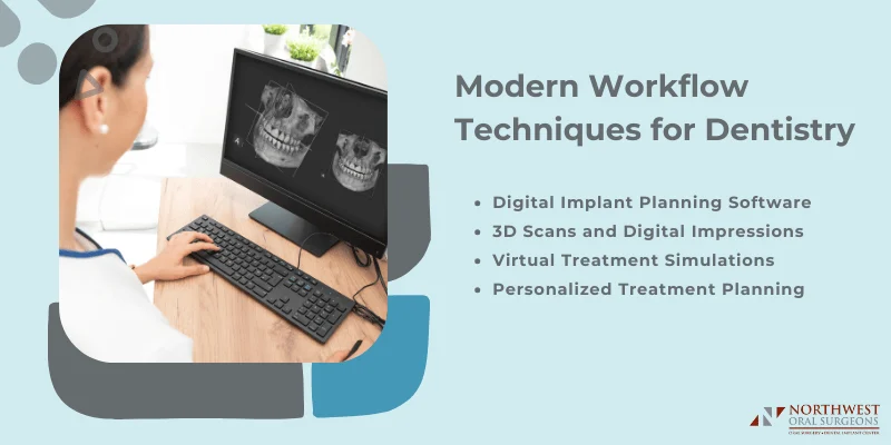
Digital Planning and Workflow Innovations
Modern digital workflows in implant dentistry integrate advanced software, 3D imaging, and virtual simulations to create precise treatment plans before surgery begins. These tools allow you and your dental team to visualize the entire procedure and identify potential problems early.
Digital Implant Planning Software
Digital implant planning software gives your dentist powerful tools to design your treatment with extreme accuracy. The software combines data from CT scans and digital impressions to create a complete view of your jaw structure, bone density, and nearby nerves.
Your dentist uses this software to determine the exact position, angle, and depth for each implant. The program can simulate different implant sizes and placements to find the best option for your specific anatomy. This level of precision wasn’t possible with traditional planning methods.
Modern implant planning software also helps your dental team coordinate with specialists and labs. Everyone involved in your care can access the same digital plan. This reduces miscommunication and ensures all professionals work toward the same goal.
The software flags potential complications before surgery starts. It alerts your dentist to areas where bone grafting might be needed or where nerves could be at risk.
3D Scans and Digital Impressions
Digital impressions replace the uncomfortable putty molds that many patients dislike. Your dentist uses an intraoral scanner to capture thousands of images of your teeth and gums in minutes. The scanner creates a precise 3D model that shows every detail.
These digital scans work with cone-beam computed tomography (CBCT) to give your dentist a complete picture. CBCT provides high-resolution 3D images of your bone structure, sinuses, and nerve pathways. When combined with digital impressions, your dentist sees both the soft tissue surface and the internal bone structure.
Digital methods are more accurate than traditional impressions. They eliminate distortion from materials setting or models being damaged during shipping. Your dentist can also retake specific areas instantly if needed without starting over completely.
Virtual Treatment Simulations
Virtual simulations let you see what your final results will look like before any work begins. Your dentist creates a digital model showing where implants will be placed and how your new teeth will function. You can view this simulation from different angles and ask questions about the process.
These simulations help your dentist practice the surgery virtually. They can test different approaches and choose the safest path. The software calculates the forces your implants will experience when you chew and ensures the placement can handle normal use.
Computer-aided design technology uses these simulations to create surgical guides. These guides fit over your existing teeth during surgery and direct the drill to the exact planned position. The guide removes guesswork and improves accuracy significantly.
Personalized Treatment Planning
Every mouth is different, and digital dentistry makes it easier to create treatment plans tailored specifically to you. Your dentist analyzes your unique bone quality, bite pattern, and aesthetic goals to design a custom approach. The digital workflow considers your medical history, lifestyle factors, and budget constraints.
Personalized treatment plans account for your individual healing patterns and risk factors. If you have conditions like diabetes or a history of gum disease, the planning software helps your dentist adjust the approach accordingly.
The plan might include additional healing time or specific implant types that work better for your situation.
Your digital plan becomes a roadmap that guides every decision throughout treatment. From the initial consultation through final restoration, each step builds on accurate digital data about your mouth.
Enhancing Surgical Precision with Guided Techniques
Modern guided techniques combine digital planning with physical surgical guides to achieve placement accuracy within 1-2 millimeters. These methods reduce human error during implant surgery and help you avoid critical anatomical structures like nerves and sinuses.
Guided Implant Surgery and Surgical Guides
Guided implant surgery uses physical templates that fit over your gums or remaining teeth to direct the drill at exact angles and depths.
These surgical guides act like a blueprint that translates your digital plan into the operating room. The latest systems achieve mean coronal deviations of just 1.11mm and angular deviations of 3.51 degrees.
You’ll find two main types of guided surgery systems available. Static guides are pre-made templates that lock the drill path in place before surgery starts. Dynamic navigation systems use real-time tracking to show the drill position on a screen as your surgeon works.
Static guides work best for straightforward cases and full-arch rehabilitation. They’re especially helpful when you need precise placement near the maxillary sinus floor or when planning sinus augmentation procedures.
Dynamic systems give your surgeon flexibility to adjust during complex cases while maintaining accuracy within 1.18mm at the implant top.
The choice between flapless and open surgery affects how your surgical guide functions. Flapless approaches using guided techniques now achieve survival rates of 98% or higher while reducing swelling and discomfort by 25-40% compared to traditional methods.
The Role of 3D Printing in Implant Placement
3D printing technology transforms your digital treatment plan into physical surgical guides within 24-48 hours. SLA (stereolithography) printers create the most accurate guides for implant placement, with tolerances under 100 microns. This precision ensures the guide fits perfectly against your teeth or gums.
Your dentist can now print guides in-house rather than waiting for lab fabrication. This cuts turnaround time by 50% and lets adjustments happen quickly if needed. The printing process uses biocompatible resins that can withstand sterilization before your surgery.
Modern digital workflows connect your CBCT scan directly to the 3D printer. The software automatically designs the guide with metal sleeves that direct the drill at the planned angle and depth. You benefit from same-day treatment planning where scanning, guide design, and printing happen in one appointment.
For complex cases involving bone grafting or multiple implants, 3D printing enables surgical guides with multiple sleeve positions. These guides maintain their accuracy even during lengthy procedures near sensitive areas like the maxillary sinus floor.
Integration of CAD/CAM in Surgical Procedures
CAD/CAM technology connects every step of your implant journey into one seamless digital workflow. The software takes your CBCT scan and intraoral scan to create a virtual model where your surgeon plans the exact implant position before touching any instruments.
Digital planning software now includes AI features that automatically identify nerves, sinuses, and bone density levels. This automation reduces planning time from 30 minutes to just 10 minutes while maintaining accuracy within 1mm. Your surgeon can simulate different implant sizes and angles to find the optimal position that avoids complications.
The CAD portion designs both your surgical guide and final restoration simultaneously. This means your crown or bridge can be pre-fabricated based on where the implant will actually sit. When immediate loading is appropriate, you leave with a temporary tooth the same day as your implant surgery.
Advanced imaging integration eliminates manual alignment steps that previously caused errors. The latest systems like NobelClinician SmartFusion automatically merge your scans without requiring registration markers. You get more predictable results because the surgical guide matches your anatomy precisely.
CAM manufacturing produces your surgical guide with metal sleeve inserts positioned at tolerances of ±5 micrometers. This level of precision proves critical during procedures near the maxillary sinus floor where even small deviations could penetrate the sinus membrane.
Predictive Analytics and Artificial Intelligence in Implantology
Advanced AI systems now analyze complex patient data to predict implant outcomes before surgery begins. These technologies use deep learning models to identify risk patterns that traditional methods might miss, helping you make more informed treatment decisions.
Deep Learning for Implant Failure Prediction
Deep learning algorithms have transformed how dentists predict implant success by analyzing large amounts of patient information at once. Convolutional neural networks (CNNs) examine your dental images, medical history, and bone quality measurements to spot warning signs of potential problems.
A neural network system developed by researchers achieved 94.48% accuracy when predicting single implant success by evaluating 55 different patient factors. These factors included your overall health, bone structure, and specific site conditions where the implant would be placed.
Another study using supervised learning methods reached 74.10% accuracy when analyzing data from 681 patients. The lower accuracy happened because the model used fewer patient factors during training. This shows why comprehensive data collection matters for reliable predictions.
Deep learning architectures can detect subtle bone changes and early signs of peri-implant disease that human eyes might miss on radiographs. Your dentist can use these predictions to adjust treatment plans before placing an implant, reducing your risk of complications later.
Evaluating Diagnostic Accuracy and Cross-Validation
Cross-validation testing ensures AI models work reliably across different patient groups. Researchers typically use five-fold cross-validation, where they split patient data into five parts and test the model multiple times to verify consistent performance.
One comprehensive evaluation tested seven different deep learning models through ten separate training cycles. The average accuracy reached about 80% across all tests, demonstrating that these tools can provide dependable predictions for your treatment outcomes.
Key performance metrics include:
- Sensitivity – how well the system catches actual problems
- Specificity – how accurately it rules out false alarms
- Accuracy – overall correctness of predictions
CNN models have shown particularly strong results in detecting peri-implant bone defects and inflammation on your radiographs. These systems outperform traditional diagnostic methods when identifying early bone level changes around implants.
The quality of training data directly affects how well these models work. Larger datasets with diverse patient information produce more accurate predictions for your specific situation.
AI-Powered Risk Assessment Tools
Modern risk assessment frameworks combine clinical, biological, and systemic data to create your personalized risk profile. These tools evaluate factors like diabetes, smoking history, and past periodontal disease to estimate your chances of developing peri-implantitis or implant failure.
Digital implant planning systems now integrate AI analytics to flag high-risk cases during the planning phase. Your dentist receives alerts about potential complications based on your bone density, implant positioning, and health conditions before surgery starts.
Some advanced platforms update your risk profile continuously during follow-up visits. By analyzing new radiographs and tissue measurements over time, these systems detect disease progression early so your dentist can intervene before serious damage occurs.
Common risk factors AI systems evaluate:
- Residual periodontal pockets
- Poor implant positioning
- Inadequate bone quality
- Systemic conditions affecting healing
These AI-driven tools don’t replace your dentist’s expertise but help them make better decisions about your care. They provide objective data analysis that supports personalized treatment strategies and preventive maintenance plans tailored to your specific risk level.
Improving Outcomes and Patient Experiences
Advanced imaging and planning technologies directly improve how quickly you heal and how satisfied you feel with your dental implant surgery. These tools help your dentist catch problems early and explain your treatment in ways that build confidence.
Reducing Recovery Times and Postoperative Risks
When your dentist uses 3D imaging for planning, you typically experience faster healing after dental implant surgery. Precise implant placement reduces surgical risks and helps your body accept titanium implants more easily.
Digital planning means less cutting and trauma to your gums during surgery. Your dentist knows exactly where to place the implant before making any incisions. This accuracy means you deal with less swelling and discomfort afterward.
Most patients return to normal activities faster with digitally planned procedures. You might need fewer pain medications and experience less bleeding. The reduced recovery time also means fewer days away from work or your regular routine.
Long-Term Stability and Early Intervention
Digital imaging helps your dentist monitor your implants over time and spot issues before they become serious. Early intervention can prevent implant failure and save you from additional procedures.
Your dentist can track marginal bone loss around your implants using the same technology. Small changes in bone levels show up clearly on digital scans. Catching these changes early allows for treatment adjustments that protect your investment.
Regular digital checkups reveal problems invisible to the naked eye. You benefit from treatment modifications before pain or mobility issues develop. This monitoring approach significantly improves your chances of keeping your implants for life.
Patient Communication and Education
Digital imaging transforms how your dentist explains your treatment plan. You can see 3D models of your jaw and understand exactly what will happen during surgery. This visual approach reduces anxiety and helps you make informed decisions.
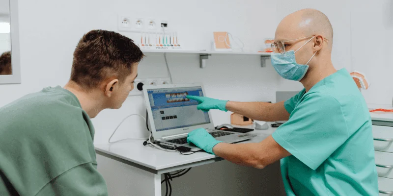
Your dentist can show you different implant placement options on screen. You see how each choice affects your final smile and bite. These clear explanations improve your patient experience and build trust in the treatment process.
Digital tools also let you preview your final results before surgery begins. You know what to expect and can request changes to the plan. This collaborative approach ensures you feel confident and satisfied with your care.
Frequently Asked Questions
Advanced imaging and careful planning play a direct role in implant success rates. The right techniques help surgeons avoid complications and place implants in the best position for long-term results.
What are the best practices for using CBCT in implant dentistry to ensure success?
CBCT scans give your surgeon a complete 3D view of your jawbone, nerves, and sinuses before surgery. This imaging shows exact bone density, height, and width so your dental team can pick the right implant size and angle.
Your surgeon should review the CBCT scan to identify where nerves and blood vessels are located. This helps avoid nerve damage that can cause numbness or tingling. The scan also shows if you need bone grafting before implant placement.
Digital planning software lets your surgeon map out the exact implant position on the CBCT images. This reduces guesswork during surgery and leads to better outcomes.
Can you explain how precise implant planning affects the outcome of dental implant surgery?
Precise planning helps your implant fuse properly with your jawbone through a process called osseointegration. When your surgeon plans the exact depth, angle, and position, the implant has the best chance to integrate successfully.
Poor planning can lead to implants placed too close to nerves or sinuses. Comprehensive pre-op evaluation and planning includes medical history review and 3D imaging to check bone quality and nearby structures.
Digital treatment planning also helps your surgeon select the right implant type for your specific bone condition. This attention to detail reduces the risk of early or late implant failure.
What are some key steps to take to prevent dental implant failure?
Managing your health conditions before surgery is critical for success. If you have diabetes, work with your doctor to control blood sugar levels because uncontrolled diabetes increases failure risk.
Quitting smoking several weeks before and after surgery greatly improves your chances of success. Smoking restricts blood flow and interferes with healing and bone integration.
Following post-operative instructions carefully protects your investment. Eat soft foods as directed, take prescribed antibiotics, and maintain gentle oral hygiene around the implant site. Keep all follow-up appointments so your dental team can catch any problems early.
Good home care after healing is just as important. Brush and floss around your implants daily and visit your dentist for regular professional cleanings.
Could you highlight the advantages of using image-guided implant planning?
Image-guided planning creates a surgical blueprint that your surgeon follows during the procedure. This technology combines CBCT scans with digital planning software to map every step before surgery begins.
Guided surgery uses custom surgical guides that fit over your gums and show exactly where to place each implant. This reduces chances of nerve or sinus injury and improves implant positioning.
The precision from image guidance means less tissue trauma during surgery. You may experience less swelling, pain, and faster healing compared to freehand placement.
Your final crown or bridge also fits better because the implant is positioned exactly where planned. This prevents mechanical problems with loose or fractured components later.
Can you describe the 3/2 rule in dental implantology and why it’s important?
The 3/2 rule is a safety guideline for implant placement in your lower jaw. It states that your surgeon should keep at least 3mm of bone above the nerve canal and 2mm of bone on each side of the implant.
This spacing protects the inferior alveolar nerve that runs through your lower jaw. Damage to this nerve causes numbness in your lip, chin, or tongue that may be temporary or permanent.
Your surgeon measures these distances on your CBCT scan during planning. If there isn’t enough space, they may recommend a shorter implant, bone grafting, or an alternative treatment plan.
Following this rule is one way experienced implant surgeons reduce the likelihood of nerve damage. Pre-surgical imaging makes it possible to maintain these safe distances.
Which imaging techniques are commonly recommended for dental implant planning?
CBCT scanning is the gold standard for implant planning today. Unlike regular dental X-rays, CBCT provides three-dimensional images that show bone in all directions.
Panoramic X-rays give a broader view of your entire jaw and can identify major obstacles. However, they don’t show bone width or provide the detail needed for precise planning.
Intraoral scans capture the exact shape of your teeth and gums. When combined with CBCT data, these scans help create surgical guides and plan your final restoration.
Some practices use digital workflows that connect all imaging types together. This gives your surgical team the most complete picture and helps them plan every detail of your treatment.