If you’re planning a dental implant, you need a strong jawbone to hold it. If your dentist finds thin, soft, or missing jawbone, a bone graft can rebuild the area so an implant will be stable and long-lasting. Knowing the signs now can save time and avoid implant failure later.
You might notice gaps, loose teeth, or a history of gum disease or long-term tooth loss that hint at bone loss. Your dentist will use imaging and exams to check bone height and density before recommending grafting or implant placement.
Key Takeaways
- Bone grafting creates enough bone to support a stable implant.
- Certain symptoms and history often indicate the need for grafting.
- Imaging and exams guide the decision and graft type.
Key Signs Bone Grafting Is Needed Before Implant Placement
You may need a bone graft when the jaw lacks the width or height to hold an implant, when disease or injury has eroded bone, or when your facial shape has changed after years without a tooth. These issues often show as sunken gums, chewing problems, or imaging that reveals thin bone.
Long-Term Tooth Loss and Bone Shrinkage
When a tooth is missing for months or years, the jawbone where the root once was loses stimulation and begins to shrink. You might notice the gum over the empty socket looks flattened or sunken. This shrinkage reduces the bone width and height needed to anchor a dental implant.
If you want an implant, your dentist will measure bone dimensions with X-rays or a 3D scan. If the bone is too thin or short, bone grafting rebuilds that area so an implant can hold firmly.
Bone graft options include your own bone, donor bone, or synthetic material. Healing time varies, but grafts often need several months before implant placement.
Jawbone Loss Due to Gum Disease or Periodontal Disease
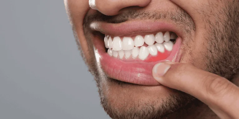
Advanced gum disease destroys the tissues that hold teeth, including the underlying bone. You may see bleeding gums, loose teeth, or pockets between teeth and gums. Over time, periodontal disease can hollow out bone where an implant would go.
Your dentist will evaluate pocket depth and bone level with clinical checks and imaging. If bone loss exists, grafting fills and supports the area, lowering the risk of implant failure.
Treating the gum disease first is important. After infection control, grafting helps restore a healthy foundation for a long-lasting dental implant.
Jaw or Facial Injury Impacting Bone Support
A sports injury, fall, or accident can break or damage the jawbone near a missing tooth. Even if the tooth was later removed, the trauma may leave areas of thin or fractured bone. You might remember the injury or notice ongoing soreness, numbness, or uneven bite.
Imaging shows where bone weakened or is irregular. Bone grafting repairs defects, fills voids, and creates solid bone for an implant to anchor into. A clinical study found 50.3% of implant patients required bone grafting to achieve adequate support.
Your surgeon may combine grafting with other repairs, and healing time depends on the size of the defect and your overall health.
Changes in Facial Structure or Sunken Cheeks
Losing several teeth or long-term missing teeth can change your facial shape. You may notice sunken cheeks, deeper wrinkles around the mouth, or a more pronounced chin. These changes happen because bone beneath the skin shrinks without tooth roots to stimulate it.
Bone grafts can restore lost bone volume and improve both function and appearance. Rebuilding the jaw supports implants and helps bring back fuller cheek contours. Your dentist can show before-and-after images or use scans to explain how grafting may affect both chewing and facial appearance.
Diagnostic Methods for Assessing Bone Quality
You need clear checks to know if your jaw can hold an implant. These checks measure bone height, bone width, and overall bone health so your dentist can plan grafting or choose an alternative.
Clinical Examination of Bone Volume
Your dentist first inspects the ridge by sight and touch. They will measure how wide and tall the ridge is with a periodontal probe or calipers and check soft tissue thickness over the bone.
A narrow, flat, or irregular ridge often means there isn’t enough bone width for the implant diameter you need. Loss of vertical height near the sinus or nerve can also appear on exam as shallow ridges or close anatomical landmarks.
You may also bite on a surgical guide or have tooth positions checked to see if the planned implant path fits available bone. This hands-on exam helps decide if a simple socket graft, a ridge-split, a block graft, or a more complex augmentation will be needed.
Role of X-Rays and CT Scans
Standard dental X-rays show general bone levels and help spot past infection or fractures. Bitewings and periapicals give a quick look at bone supporting adjacent teeth and at vertical height. They do not measure width well.
A medical CT gives 3D detail about bone height, width, and internal structure when X-rays are unclear. CT shows the sinus floor, nerve canal, and extent of bone loss. Your clinician may order a CT when complex anatomy or major grafting is likely.
If you already have an X-ray, expect the dentist to compare it with clinical findings to confirm whether bone volume looks sufficient for the implant you plan.
CBCT Scan for Bone Height and Width
A cone-beam CT (CBCT) gives high-resolution 3D images focused on the jaws. It measures bone height and bone width in millimeters at the exact implant site and shows the relationship to the maxillary sinus and inferior alveolar nerve.
CBCT also helps the dentist plan implant diameter and length and decide whether a simultaneous graft-and-implant or a staged graft is safer.
Your clinician will review axial, coronal, and sagittal slices and may use virtual implant planning software to test implant positions. This reduces surprises during surgery and clarifies if a dental bone graft is required to reach the dimensions needed for stable implant placement.
Not sure if you have enough bone for implants? Book a personalized evaluation now.
Common Causes Leading to Bone Grafting Before Implants
You may need grafting when your jaw no longer has enough height or width to hold an implant. Damage can come from long-term denture wear, birth defects that leave thin bone, or problems at a past extraction or implant site.
Prolonged Denture Use and Ridge Resorption
Wearing removable dentures for years often leads to ridge resorption. The denture presses on the jaw and the bone loses stimulation. Over time the bone shrinks in height and width, so your jaw becomes too narrow for a stable implant.
That loss changes how your face looks and how dentures fit. You might notice loose dentures, difficulty chewing, or a sunken look around the mouth. A ridge augmentation or bone graft can rebuild the lost bone so an implant will be stable.
Your dentist will measure bone with x-rays or a 3D scan. If the ridge is under the necessary width or height, they’ll recommend graft material and a plan for grafting before implant placement.
Congenital Bone Deficiencies
Some people are born with thin or missing jawbone in one or both arches. This can come from genetic conditions or simply from the way the jaw developed. You might never have had a tooth in that spot or you may have an asymmetric smile.
Congenital bone issues can make implant placement risky without grafting. Your clinician will assess the defect size and suggest a targeted bone graft or ridge augmentation. They may use your own bone, donated bone, or synthetic material to build volume.
Planning is important when the defect is large. Staged grafting helps create a predictable base for future implants and improves the long-term success of the restorations.
Failed Previous Implant or Extraction Site Complications
When an implant fails or an extraction site becomes infected, bone loss often follows. Infection, chronic inflammation, or improper healing can leave holes or thin bone where a tooth once was. That damaged area may not hold a new implant safely.
You may have symptoms like pain, swelling, or a persistent gum pocket. Imaging will show the defect size. Treatment commonly includes cleaning the site, removing infected tissue, and placing a bone graft to fill the void.
Sometimes grafting happens at the same time as implant replacement if the defect is small. For larger defects, your provider will perform ridge augmentation and wait for healing before placing the implant.
Types of Bone Grafts and Grafting Procedures for Implant Preparation
You’ll learn which graft choices fit different problems and what each procedure does to prepare your jaw for an implant. Expect clear options, typical healing time, and what each graft aims to fix.

Overview of Types of Bone Grafts
You can get bone from four main sources: your own body (autograft), a human donor (allograft), an animal source like bovine (xenograft), or synthetic materials (alloplast).
Autografts often heal fastest because they carry your living cells, but they require a second surgical site; commonly the chin or hip. Allografts and xenografts avoid that second site and act as a scaffold for your bone to grow into. Synthetic grafts can be shaped precisely and avoid biological risks.
Key choices depend on how much bone you need, where the defect sits, and your health. Your dentist will weigh healing speed, infection risk, and whether you need structural support now or just a scaffold for later bone growth. Typical integration time before implant placement ranges from 3 to 6 months, sometimes longer for large grafts.
Sinus Lift for Upper Jawbone Support
A sinus lift raises the sinus floor and places graft material under the sinus membrane when your upper jaw lacks height.
This matters most in the back upper teeth where the sinus can expand after tooth loss or with long-term bone resorption. Your clinician opens a small window in the lateral sinus wall or uses a crestal approach, lifts the membrane gently, and packs graft material; often a xenograft or allograft; into the new space.
You usually heal for 4–9 months before an implant is placed, depending on graft size and material. Risks include sinus membrane tear and infection; your provider will evaluate sinus health with imaging first. Smoking and sinus issues can slow healing and may change the treatment plan.
Ridge Augmentation to Increase Bone Width
Ridge augmentation builds width or shape when your jaw is too narrow to hold an implant safely.
The surgeon exposes the ridge, adds graft material to the deficient area, and often uses a barrier membrane to keep soft tissue out while bone grows in. Materials vary: autograft for strong structural support, or allograft/xenograft and synthetic mixes when you want to avoid a donor site.
This procedure is common after long-term tooth loss or trauma that left the ridge flat. Healing usually takes 3–6 months; larger defects may need staged grafting before implant placement. Your dentist will check soft tissue thickness too, since thin gums can expose graft material and increase complications.
Benefits of Bone Grafting for Dental Implant Success
Bone grafting builds the jaw so implants sit in strong, healthy bone. It helps the implant fuse to your jaw, keeps bone from shrinking, and improves how your smile looks and works.

Enhanced Osseointegration and Implant Stability
A bone graft gives your implant the solid bone surface it needs for osseointegration, the direct bonding of bone to the implant metal.
When grafted bone reaches good density, the implant threads can contact living bone across more surface area. That reduces micromovement during healing and raises the chance the implant stays secure.
Your dentist will often wait until scans show enough bone volume before placing the implant. In some cases they place the graft and implant together; in others they stage the graft first and place the implant after several months.
Either way, grafting improves primary stability at placement and long-term mechanical support for chewing forces.
Preventing Additional Bone Loss
When a tooth is missing, the jawbone naturally shrinks where the tooth root used to stimulate it. A bone graft restores lost volume and fills gaps left by extractions, infections, or disease. This stops the collapse of nearby bone and supports surrounding teeth and tissues.
By restoring height and width, grafting also prevents the sinus from expanding into implant sites in the upper jaw. That avoids future complications and the need for more extensive surgery later. Keeping bone volume now reduces the chance you’ll need larger corrective grafts later on.
Long-Term Aesthetic and Functional Outcomes
A stable bone foundation keeps the implant crown aligned with your bite and adjacent teeth. That preserves chewing efficiency and prevents uneven wear. Proper bone shape also supports the gum contour, so your smile looks natural and the prosthetic tooth matches the gum line.
Grafting lowers the risk of implant failure, loosening, or sunken soft tissue that can reveal metal or make a tooth look short. It also helps maintain facial structure by preventing bone collapse that can change jawline and lip support. These benefits combine to give you a predictable, durable result for years.
Frequently Asked Questions
This section answers clear questions about when bone grafting is needed, how clinicians check your jaw, and what signs to watch for. It explains causes, the role of bone density, and the “3/2 rule” so you know what to expect before implant surgery.
What indicators suggest bone grafting might be necessary for dental implants?
Your dentist will look for low jaw height or width, thin ridge shape, and areas where bone has resorbed after tooth loss. They will also note prior infections, trauma, or long-term tooth absence that reduced bone volume.
Imaging that shows insufficient bone to fully surround an implant is a direct indicator. If the implant would lack at least 1–2 mm of bone on its sides, grafting is often recommended.
How do dentists evaluate the jawbone for implant placement?
Your dentist uses dental X-rays and a CBCT scan to measure bone thickness, height, and quality precisely.
These images show sinus position, nerve location, and any bone defects that affect implant placement.
They combine imaging with a clinical exam of the gums and past dental history. That helps create a treatment plan and decide if a graft, sinus lift, or alternative is needed.
Can you explain what the 3/2 rule means for implant success?
The 3/2 rule is a guideline about bone coverage and implant length. It suggests you want at least 2 mm of bone on the buccal (cheek) side and 3 mm of total surrounding bone to help long-term stability.
This rule helps clinicians judge whether an implant will be stable without grafting. If measurements fall short, your dentist may recommend bone grafting to meet those minimums.
Are there specific signs that I might see if my jawbone is inadequate for implants?
You may notice a sunken cheek, shifting teeth, or a change in bite where missing teeth were. Receding gums or loose adjacent teeth can also signal underlying bone loss.
Often you won’t feel pain from low bone; imaging is the reliable way to detect the problem. Ask for a CBCT scan if you suspect hidden bone loss.
What are common reasons a patient would need a bone graft before getting implants?
Long-term tooth loss without replacement allows the jawbone to shrink. Severe periodontal (gum) disease and past infections can destroy supporting bone, too.
Trauma, congenital thin bone, or an enlarged sinus cavity near upper molars also create the need for grafting. Failed extractions that left poor healing spots can require graft repair before implants.
How does bone density affect the implant process, and why is grafting sometimes required?
Higher bone density gives implants better primary stability and faster integration. Low-density or porous bone increases the risk of implant movement and failure during healing.
Grafting adds volume or improves bone quality so the implant has enough support. In the upper back jaw, a sinus lift graft raises bone height to allow proper implant length.